The ability of continuous cell lines to exist almost indefinitely in vitro has opened the possibility of questionable subculturing practices and hence, questionable scientific data. The degree of subculturing a cell line has undergone is often expressed as “passage number,” which can generally be thought of as the number of times cells have been transferred from vessel-to-vessel. A growing body of literature demonstrates passage number affects a cell line’s characteristics over time.1-6 Cell lines at high passage numbers experience alterations in morphology, response to stimuli, growth rates, protein expression, and transfection efficiency, compared to lower passage cells.
The scientific community is taking notice that cell line quality is crucial to successful experimentation and that avoiding the use of cell lines that have been in culture too long is an important step to ensure reliable and reproducible results. But while the evidence for passage number-related effects on cell lines is compelling, much less is understood about the mechanisms underlying passage dependent changes and about actions researchers can take to avoid passage number effects in their experiments.
Compelling data for passage effects
Studies examining passage-dependent effects on mammalian cell lines are varied, focusing on a wide range of cell types and functions such as tumorigenicity, differentiation, gene expression and cell signaling. Passage number can affect insect cell lines as well, significantly decreasing protein yields and cellular productivity.7,8,9 The following examples from peer-reviewed literature center on the differences between low and high passage cell lines.
Stably-transformed mouse insulinoma cells
A study by O’Driscoll compared MIN-6 cells at low passage (passage 18) and high passage (passage 40) and found significant differences in the expression of numerous mRNAs involved in regulated secretion, adhesion, and proliferation.10 With almost 1,000 genes discovered to be differentially expressed between low and high passage cells, their study also suggests passage number plays a role in the differentiation state of this cell type.
Human prostate cells
The common prostate cancer cell line LNCaP was examined at low (passage 25) and high (passage 60) passage in another study by Lin.11 The analysis here demonstrated that the PI3K/Akt pathway regulated androgen receptor activity in a passage number-dependent manner which may have implications in the various stages of prostate cancer.
By beginning with the concept that cells in culture are under environmental and manipulative stress, both the “why” and “how” genotypic and phenotypic changes that occur become more apparent, including the mechanisms that underlie passage effects. Cells in culture are continually subjected to the evolutionary processes of competition and natural selection.
Most cell cultures represent heterogeneous populations that compete for resources such as growth factors, salts, and nucleic acids. When given an advantage, such as a faster growth rate, one cell type may overgrow another within a single population. Such competition gives rise to extant populations that no longer correctly represent the original starting material. Events such as dedifferentiation and loss of tissue-specific function should be considered the norm as passage numbers increase.
Transformed and diseased cell lines are of special concern, since they represent abnormal starting populations in which evolutionary changes occur rapidly at both the genotypic and phenotypic levels over time. In these cell types one or all of the typical cellular checkpoint genes, such as p16/INK4a, pRB and p53, have been altered whereby the cells have become “immortal.” These alterations are often in parallel with other cellular mutations, and the continual subculture of these cell lines exacerbates genomic instability.
How many passages are too many?
A straightforward method for determining the passage number of a cell line does not exist. A review of the literature on passage-related effects in cell lines demonstrates that the effects are complex and heavily dependent on a host of factors such as the type of cell line, the tissue and species of origin, the culture conditions and the application for which the cells are used. For example, unpublished data at ATCC show high-passage Caco-2 cells exhibit an increase in the expression of GFP reporter gene after transfection, while high passage MCF7 cell lines exhibit a decrease in GFP levels.
Further complicating matters is that a passage level considered “high” for one cell line may not give rise to any significant passage effects in another. Preventing passage-related effects from influencing experiments becomes a matter of determining the passage number range under which a set of experiments can be consistently performed for a given cell line.
Essential first steps to minimizing cell line passage effects
Researchers have a variety of weapons at their disposal to help avoid passage-dependent effects and ensure valid and reproducible experimental results. Starting with high-quality cells is an essential first step as cells from well-known biological resource centers (BRCs) are likely to be well characterized, more extensively tested, and from lower passages. For example, ATCC follows a strict seed-stock cellbanking method to ensure distribution of consistent, low-passage cell culture.12
Optimizing the cells’ environment is also key—carefully select media and sera, control pH with buffers and gases, monitor temperature and pay attention to the growth surface.13 Practice good cell culture technique by harvesting, feeding, and storing cells according to growth curve data.13,14 Finally, conduct authentication and characterization testing on cell lines derived locally or obtained from sources other than BRCs.
Assess and minimize passage number levels
When working with cell lines, it is good cell culture practice to conduct fundamental tests to designate an acceptable passage number range that maintains consistent cell performance.
Researchers should establish baselines and reference points for use in checking for unacceptable differences in experiments or applications. Routine cell line monitoring includes cell morphology checks, identifying markers for genes of interest and correlating expression with passage number, as well as establishing experimental criteria such as growth rates or protein expression levels.
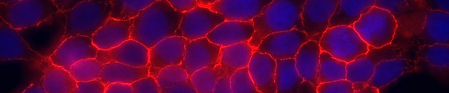
Observing cellular morphology is the simplest and most direct method used to identify the health and stability of cells. Obtaining morphology information from comparative observations both at high and low densities of cultures depends on knowledge of several factors. Morphology can vary between lines depending on the health of the cells and, in some cases, the differentiation state. Morphology can change with plating density as well as with different media and sera combinations. Cell morphology is best monitored through frequent, brief observations. In general, if a culture has an unusual appearance, there is likely a problem. It is recommended that researchers be alert during periodic morphology checks and maintain cell morphology images for comparisons.
Evaluating rates of cell proliferation from growth curve data can yield valuable information about a culture’s response to a stimulus. Sudden decreases or increases are a sign that changes may have occurred within the population. Performing a growth curve analysis is a crucial element for monitoring the consistency of the culture and determining a number of other key characteristics, such as the best time to subculture, the optimum dilution and the estimated plating efficiency at various cell densities. Growth curve analysis can also help determine population doubling times and should be performed routinely when enzymatic or functional analysis is imminent. Using cell lines with consistent growth properties should be pursued as a rule.
Download a PDF of this technical document
Download NowReferences
- Esquenet M et al. LNCaP prostatic adenocarcinoma cells derived from high and low passage numbers display divergent responses not only to androgens but also to retinoids. Journal of Steroid Biochemistry and Molecular Biology. 62:391-399, 1997.
- Briske-Anderson MJ et al. Influence of culture time and passage number on morphological and physiological development of Caco-2 cells. Proceedings of the Society for Experimental Biology and Medicine. 214(3): 248-257, 1997.
- Chang-Liu CM et al. Effect of passage number on cellular response to DNA-damaging agents: cell survival and gene expression. Cancer Letters. 26(113):77-86, 1997.
- Yu H et al. Evidence for diminished functional expression of intestinal transporters in Caco-2 cell monolayers at high passages. Pharmaceutical Research. 14(6): 757-762, 1997.
- Wenger SL et al. Comparison of established cell lines at different passages by karyotype and comparative genomic hybridization. Bioscience Reports. 24(6): 631-639, 2004.
- Sambuy Y et al. The Caco-2 cell line as a model of the intestinal barrier; influence of cell and culture related factors on Caco-2 cell functional characteristics. Cell Biology and Toxicology. 21: 1-26, 2005.
- Calles K et al. Effects of Conditioned Medium Factors and Passage Number on Sf9 Cell Physiology and Productivity. Biotechnology Progress. 22: 394-400, 2006.
- Joosten CE et al. Effect of culture conditions on the degree of sialylation of a recombinant glycoprotein expressed in insect cells. Biotechnology Progress. 19: 739-749, 2003.
- Clemm DL. Scale-up of protein production in a stirred bioreacto. In Baculo Virus Expression Vectors: A Laboratory Manual; O’Reilly, D. R., Miller, L. K., Luckow, V. A., Eds.; W. H. Freeman: New York; pp 241-248, 1992.
- O’Driscoll L et al. Phenotypic and global gene espression changes in low and high passage MIN6 cells. J of Endocrinology. 191: 665-676, 2006.
- Lin H et al. Suppression Versus Induction of Androgen Receptor Functions by the Phosphatidylinositol 3-Kinase/Akt Pathway in Prostate Cancer LNCaP Cells with Different Passage Numbers. Journal of Biological Chemistry. 51: 50902-50907, 2003.
- ATCC. Maintaining high standards in cell culture. Brochure. 2010.
- Hartung T et al. Good cell culture practice: ECVAM good cell culture practice task force report 1. ATLA 30: 407-414, 2002.
- Hay RJ et al. Cell Line Preservation and Authentication. In: “Animal Cell Culture.” JRW Masters (ed.). J. Wiley, Inc., Oxford University Press. New York City. 2000.
Further reading
- Riley SA et al. Active hexose transport across cultured human Caco-2 cells: characterization and influence of culture conditions. Biochimica et Biophysica acta. 1066(2): 175-182, 1991.
- Langeler EG et al. Effect of culture conditions on androgen sensitivity of the human prostate cancer cell line LNCaP. Prostate. 23(3): 213-223, 1993.
- MacLeod RA et al. Identity of original and late passage Dami megakaryocytes with HEL erythroleukemia cells shown by combined cytogenetics and DNA fingerprinting. Leukemia. 11(12): 2032-2038, 1997.
- Vierck JL et al. Interpretation of cell culture phenomena. Methods in Cell Science. 22(1): 79-81, 2000.
- Behrens I et al. Do cell culture conditions influence the carrier mediated transport of peptides in Caco-2 cell monolayers? European Journal of Pharmaceutical Sciences. 19(5): 433-442, 2003.