
2021 Photo Contest – Winners Announced
Congratulations to the winners
Thank you to all of the scientists who submitted images featuring their innovative research and scientific discoveries using ATCC cell lines or microbial strains. We are pleased to announce the winners of our 2021 Photo Contest!
After receiving over 17 thousand votes through the contest, we have identified the top 2 most popular photographs for Cell Biology and Microbiology. Further, through careful deliberation by an awards committee at ATCC, we have identified the top 5 photographs in the scientific excellence category. Check out the winning images below!
2021 Most Popular Photograph Awards
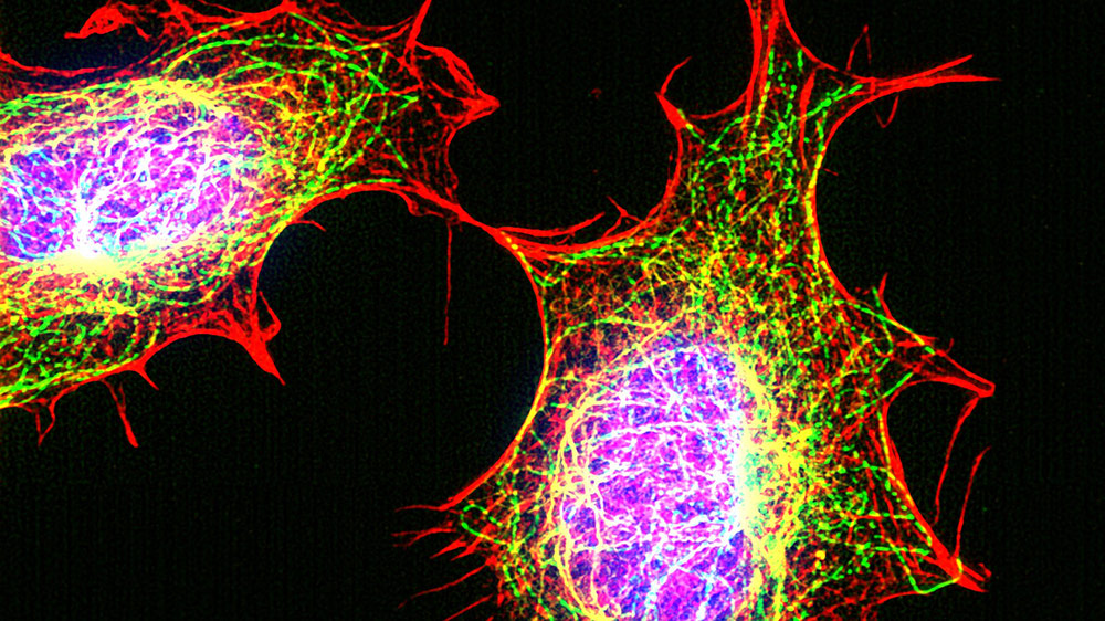
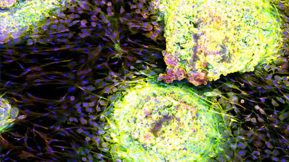
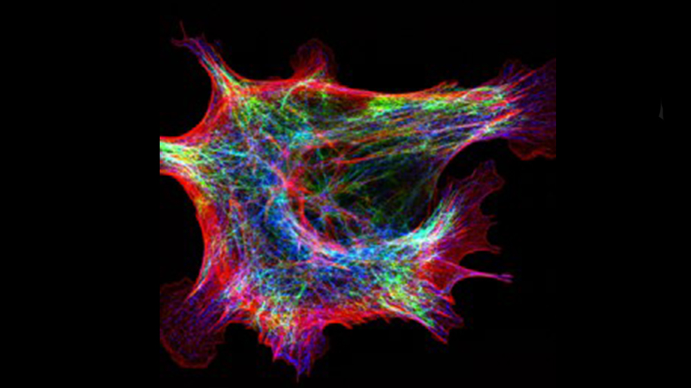
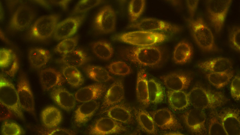
Photographer - Yuan Yuan, Harvard University
NIH/3T3 cells (ATCC CRL-1658) were fixed with 4% formaldehyde in PBS. Microtubule (green) and Vimentin (yellow) are stained with antibodies. Actin (red) is stained with phalloidin-TRITC. Nucleus (blue) is stained with DAPI. Cells were imaged by structure illumination microscopy.
"ATCC provides me cell lines that make my research reliable and repeatable."
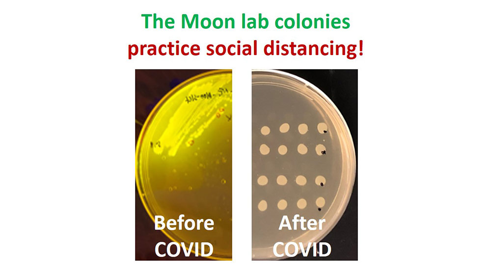
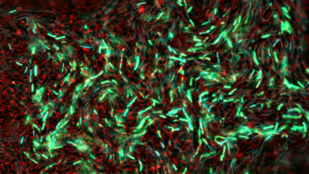
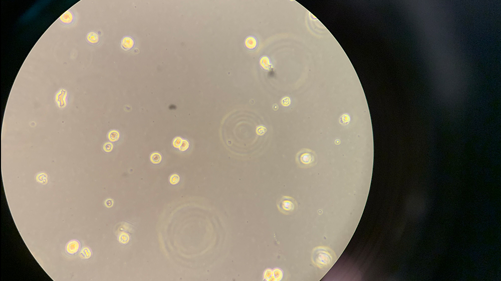
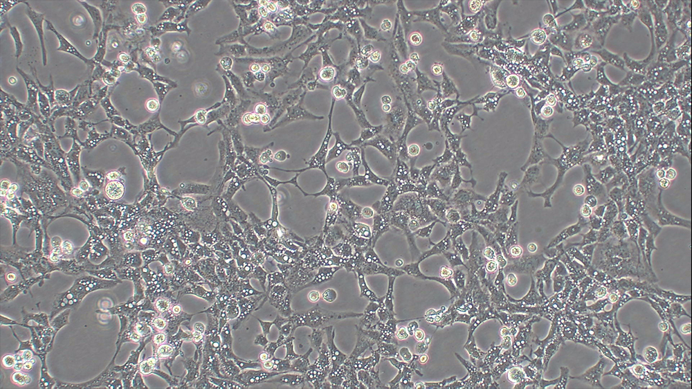
Photographer - Tae Seok Moon, Washington University in St. Louis.
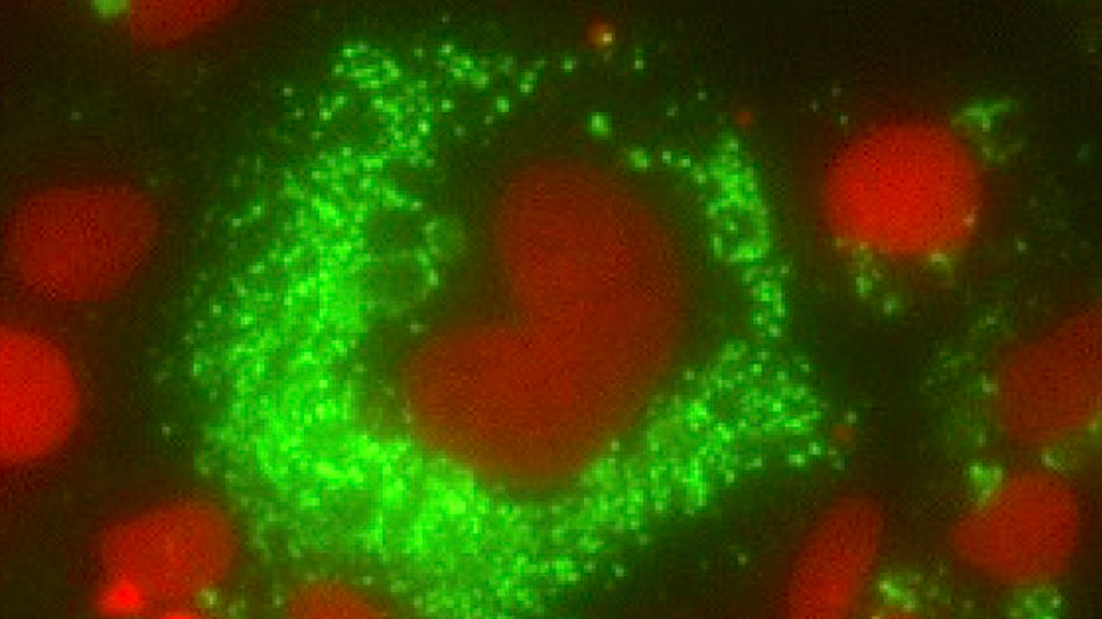
Salmonella enterica subsp. enterica serovar Typhimurium (ATCC 53647) expressing GFP (left panel) and not expressing GFP (right panel).
"Salmonella typhimurium (BSL1 strain) has a genotype/phenotype similar to that of the pathogenic version."
2021 ATCC Excellence Photograph Awards
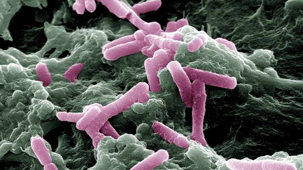
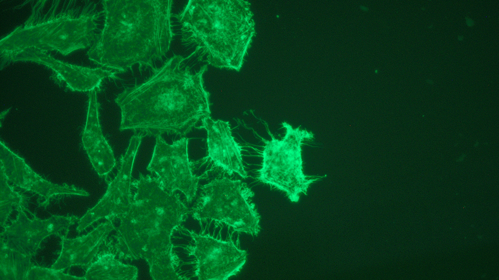
Photographer - Mindy Engevik, Medical University of South Carolina
Scanning Electron Microscopy image of Clostridioides difficile R20291 (pink) binding to LS 174T cells (ATCC CL-188) (green).
"ATCC provides valuable cell lines and microbes to examine bacterial host interactions."
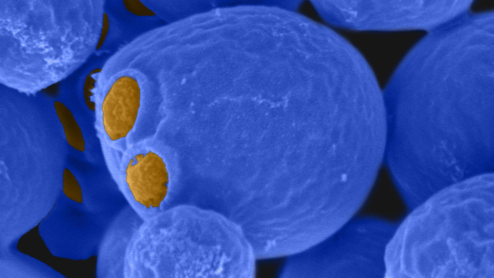
Photographer – Matthew Winans, West Virginia University
Scanning Electron Micrograph of Saccharomyces cerevisiae strain BY4741 (ATCC 4040002)
"The strain and species selection of ATCC has allowed my research to progress in areas of life sciences beyond what was normal in my lab."
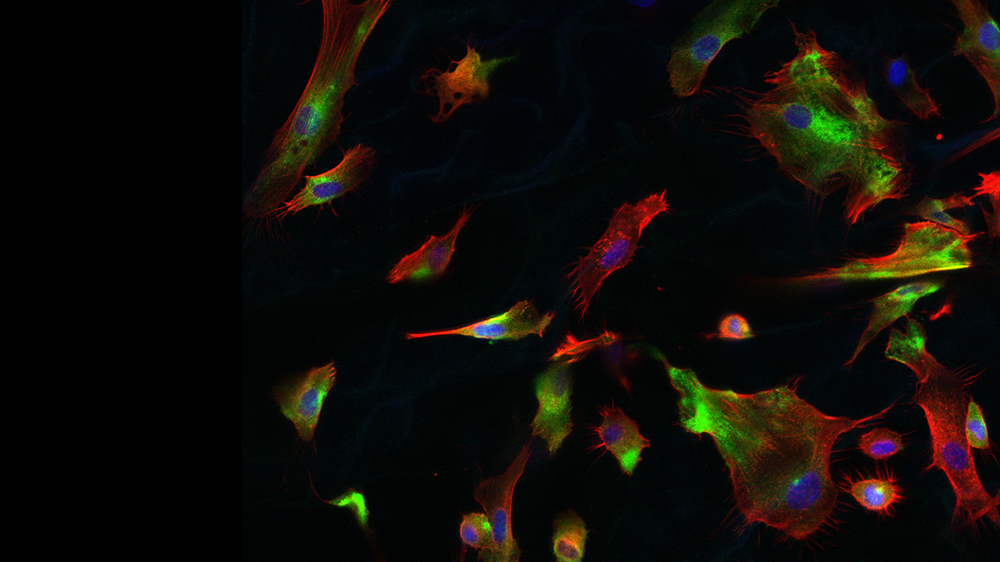
Photographer - Alexandra Paul, University of Texas at Austin
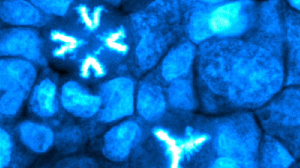
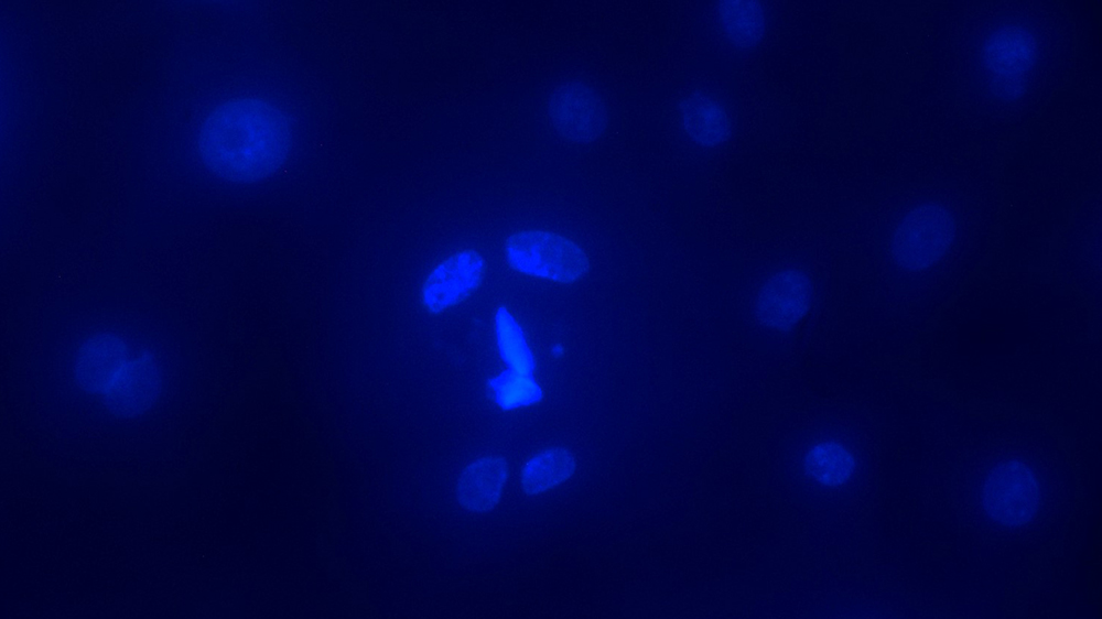
Three different stages of cell division are visible next to each other. The Calu-3 cells (ATCC HTB-55) are stained with DRAQ5 and imaged with confocal laser scanning microscopy. The condensed DNA in dividing nuclei appears brighter than non-dividing nuclei.
"We purchased Calu-3 cells to study the biomechanics behind SARS-CoV-2 infections. Using this cell line, we built different models for young and aged lungs."
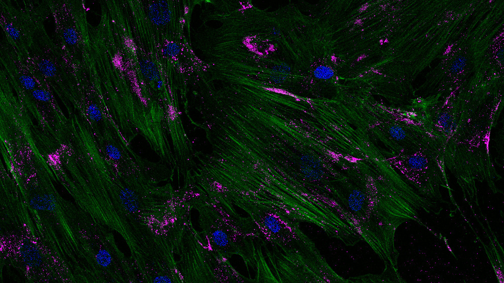
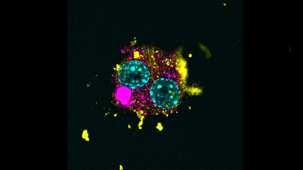
Photographer – Elisabet Olsen, Tulane University
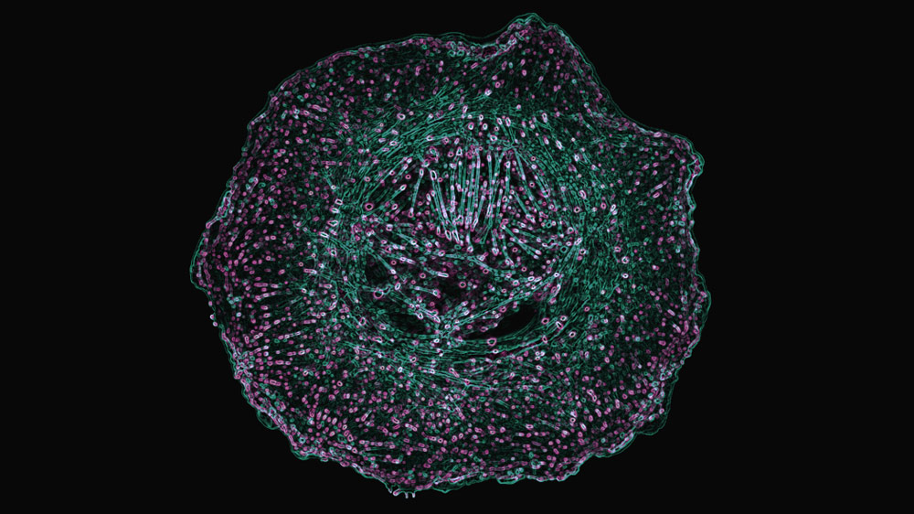
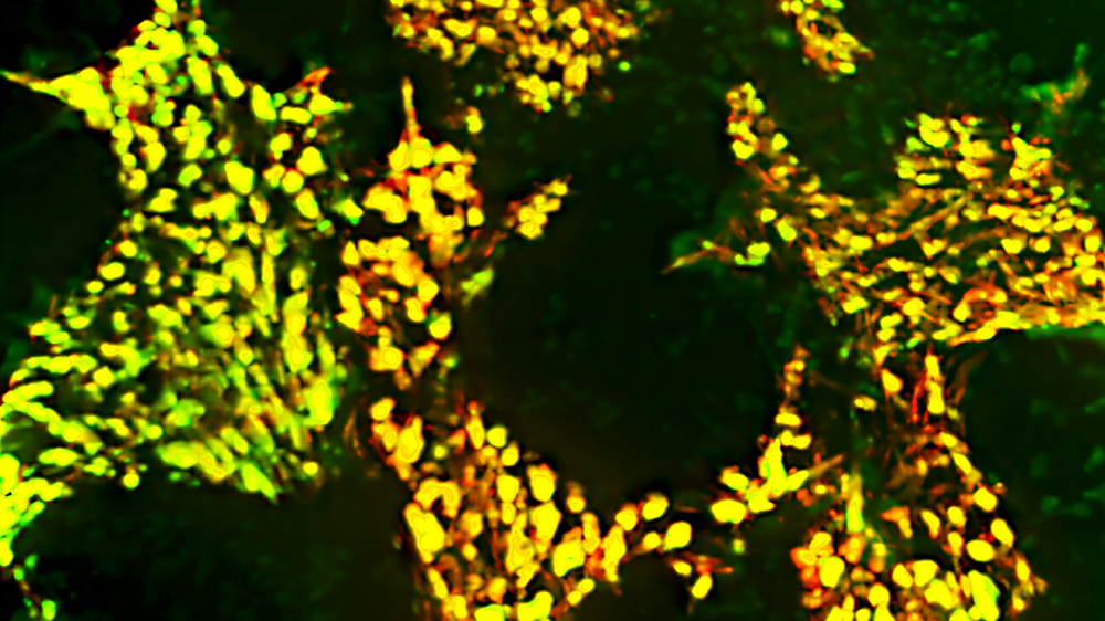
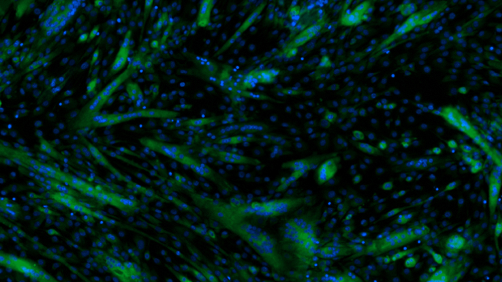
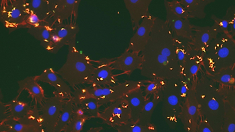
Engineered 3D lung tumor microenvironment. Non-small cell lung adenocarcinoma cells (ATCC CCL-185) grown as spheroid were embedded in 3D collagen gels with lung cancer-associated fibroblasts. Hydrogels were fixed and stained for dapi (blue), smooth muscle actin (green), and activated Yes-associated protein (YAP) 1 (red). 3D tumor invasion is observed.
"I obtain my cells and growth media from ATCC and without their service I would have a hard time conducting my research. Most importantly, ATCC has the best customer service I have ever experienced!"
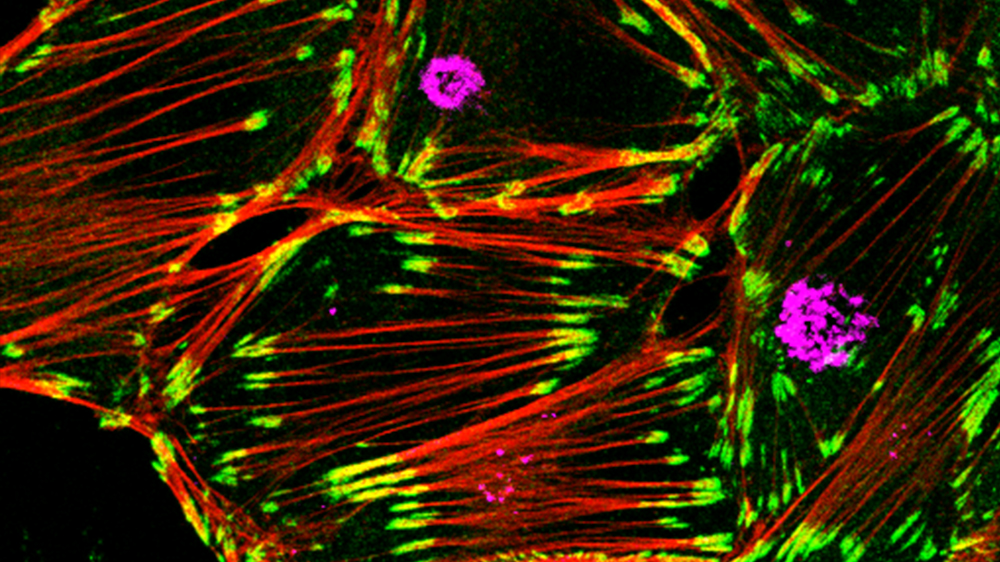
Photographer – Tejeshwar, Rao, University of Alabama at Birmingham
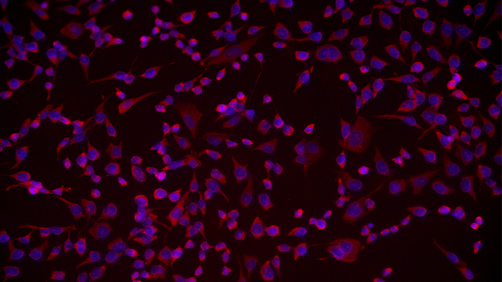
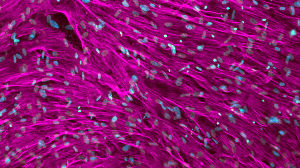
Primary Human Umbilical Vein Endothelial Cell (ATCC PCS-100-013) depicting the radiant distribution of actin cytoskeleton (green) and cell contact points (focal adhesions) represented via paxillin staining (magenta). The cells were imaged using Total Internal Reflection Fluorescence Microscopy.
"All cell lines used in our laboratory are directly purchased via ATCC. Media and supplementary additives necessary for cell culture are also purchased from ATCC. We always refer to the associated protocols available on your website for our culturing and freezing needs."
View all the contest entries
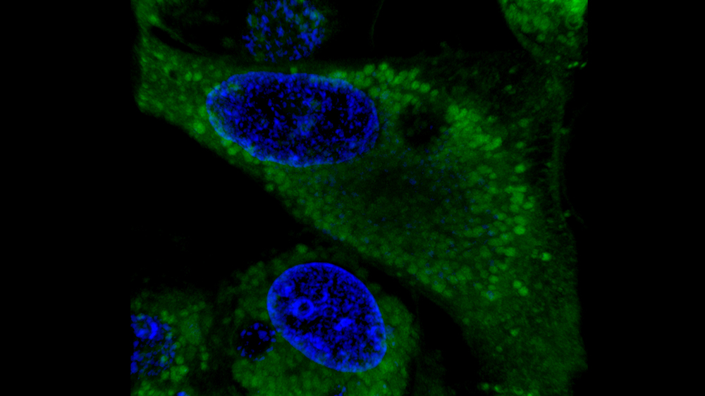
Photographer - Mrinmoyee Majumder, MUSC
Oncogenic protein's cytoplasmic expression in A549 cells (ATCC CCL-185)
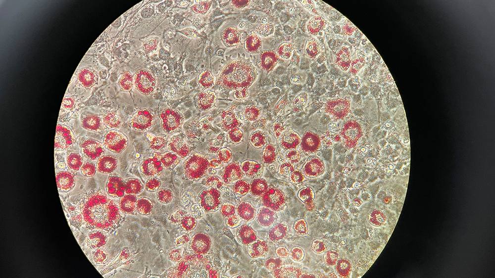
Photographer - Paola Munoz Tello, The Scripps Research Institute
3T3-L1 (ATCC CL-173) red oil staining after differentiation
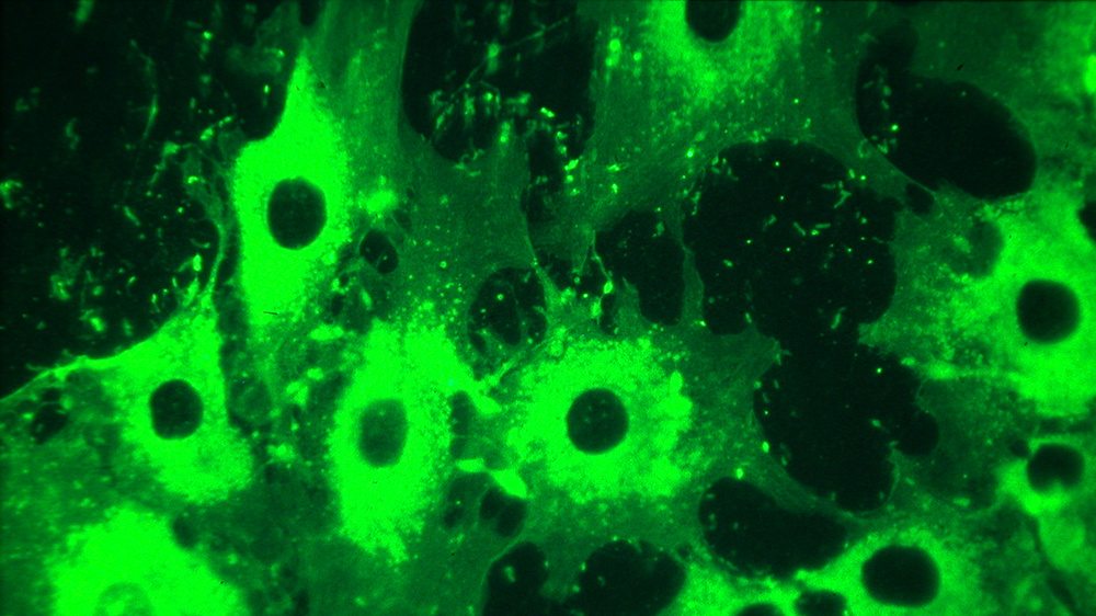
Photographer - Yihan Zhang, New Jersey Institute of Technology
Escherichia coli GFP (ATCC 25922GFP) was cultured with ATCC Medium 2855 and used as the bacterial inoculum to simulate the biofilm formation, then the coverslips were visualized with an epifluorescence microscope for bacterial attachment with or without Nanobubbles treatment.
2021 ATCC Excellence Photograph Award
Photographer - Alexandra Paul, University of Texas at Austin
Three different stages of cell division are visible next to each other. The Calu-3 cells (ATCC HTB-55) are stained with DRAQ5 and imaged with confocal laser scanning microscopy. The condensed DNA in dividing nuclei appears brighter than non-dividing nuclei.
2021 ATCC Excellence Photograph Award
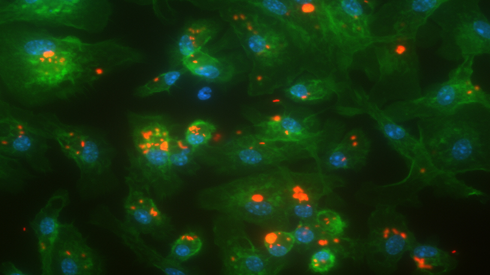
Photographer – Elisabet Olsen, Tulane University
Engineered 3D lung tumor microenvironment. Non-small cell lung adenocarcinoma cells (ATCC CCL-185) grown as spheroid were embedded in 3D collagen gels with lung cancer-associated fibroblasts. Hydrogels were fixed and stained for dapi (blue), smooth muscle actin (green), and activated Yes-associated protein (YAP) 1 (red). 3D tumor invasion is observed.
2021 ATCC Excellence Photograph Award
Photographer – Tejeshwar, Rao, University of Alabama at Birmingham
Primary Human Umbilical Vein Endothelial Cell (ATCC PCS-100-013) depicting the radiant distribution of actin cytoskeleton (green) and cell contact points (focal adhesions) represented via paxillin staining (magenta). The cells were imaged using Total Internal Reflection Fluorescence Microscopy.
2021 Most Popular Photograph Award
Photographer - Yuan Yuan, Harvard University
NIH/3T3 cells (ATCC CRL-1658) were fixed with 4% formaldehyde in PBS. Microtubule (green) and Vimentin (yellow) are stained with antibodies. Actin (red) is stained with phalloidin-TRITC. Nucleus (blue) is stained with DAPI. Cells were imaged by structure illumination microscopy.
2021 ATCC Excellence Photograph Award
Photographer – Matthew Winans, West Virginia University
Scanning Electron Micrograph of Saccharomyces cerevisiae strain BY4741 (ATCC 4040002)
2021 ATCC Excellence Photograph Award
Photographer - Mindy Engevik, Medical University of South Carolina
Scanning Electron Microscopy image of Clostridioides difficile R20291 (pink) binding to LS 174T cells (ATCC CL-188) (green).
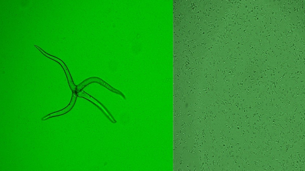
2021 Most Popular Photograph Award
Photographer - Tae Seok Moon, Washington University in St. Louis.
Salmonella enterica subsp. enterica serovar Typhimurium (ATCC 53647) expressing GFP (left panel) and not expressing GFP (right panel).
Photographer – Kristen Engevik, Baylor College of Medicine
Bone marrow-derived macrophages marked with phallodin (pink) and Hoechst (yellow) engulfing E. coli K12 (ATCC PTA-755) (blue).
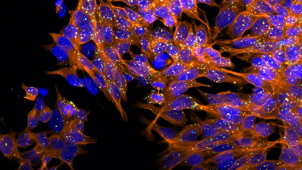
Photographer – Jillian Eskra, Johns Hopkins University
Individual RNA transcripts for POLR2A (yellow) and PPIB (light blue) expressed by 22Rv1 prostate cancer cells (ATCC CRL-2505) can be visualized as single spots using RNA in situ hybridization. Microtubules are displayed in orange.
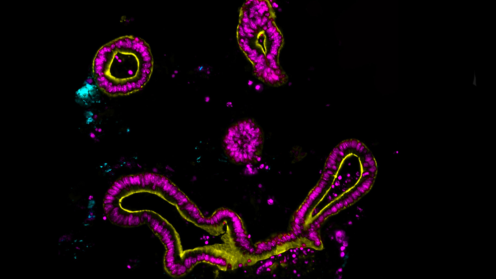
Photographer – Taylor Ticer, Medical University of South Carolina
Mouse jejunal organoids incubated with fluorescently tagged Acinetobacter calcoaceticus (ATCC 23055)
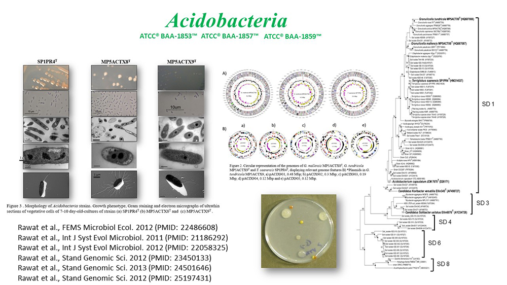
Photographer – Suman Rawat, Rutgers University
Cold-adapted Acidobacteria from Arctic tundra. 1. Terriglobus saanensis (ATCC BAA-1853); 2. Granulicella mallensis (ATCC BAA-1857); 3. Granulicella tundricola (ATCC BAA-1859)
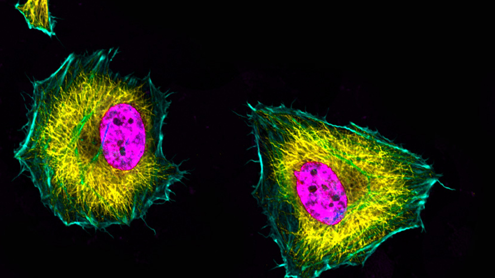
Photographer – Amy Engevik, MUSC
Hela cells (ATCC CCL-2) stained for alpha tubulin in yellow, f-actin in teal and nuclei in pink.
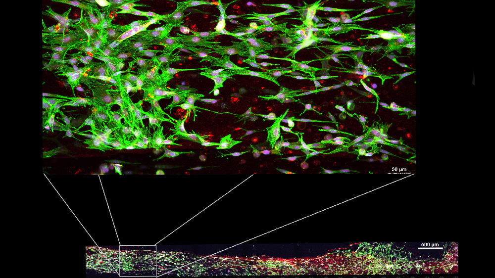
Photographer – Melanie Hilman, University of Pennsylvania
Growing human skeletal myocytes (ATCC PCS-950-010) in microchannels.
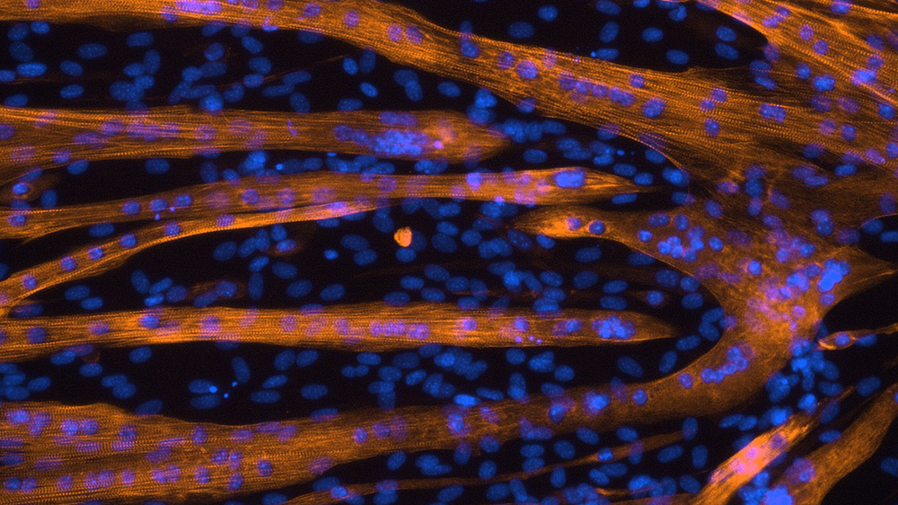
Photographer – Spencer Miller, Indiana University Medical School
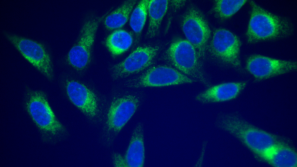
10x fluorescent image of C2C12 myotubes (ATCC CRL-1772) grown in plastic 6-well culture plates and fixed with 4% paraformaldehyde in PBS. Myosin heavy chain (all isoforms; orange) was stained using antibodies. Nuclei (blue) were stained using DAPI.
Photographer – Heli Modi, Integra Life Science
In wound healing, fibroblasts migrate, proliferate, and then secrete extracellular matrix (ECM) proteins into the wound bed. ATCC adult fibroblast cells (ATCC PCS-201-012) were used to evaluate cell proliferation on Duragen matrix. Integra's Duragen matrix is one of the safest and most effective onlay grafts for the restoration and repair of dura mater. Cells were stained for F-actin (red), Focal contacts (green) and cell nucleus (blue).
Photographer – Korinn Murphy, UNMC
The obligate intracellular pathogen Chlamydia trachomatis infecting a FAK knock-out fibroblast cell (ATCC CRL-2644). Sample is stained for chlamydia (magenta) actin (red) and paxillin (green).
Photographer – Sandra Story, NUBAD LLC
Bacteria inspired by Van Gogh. Bacterial cytological profiling for the discovery of new drugs to combat multidrug-resistant bacteria. Fluorescence image of Bacillus subtilis (ATCC 6051) stained with SynaptoRed, SYTOX Green and DAPI after treatment with ciprofloxacin.
Photographer – Jing Zia, Harvard University
Osteogenic differentiated hBMSC cells (ATCC PCS-500-012) stained with myosin IIA (purple) and nuclei (blue).
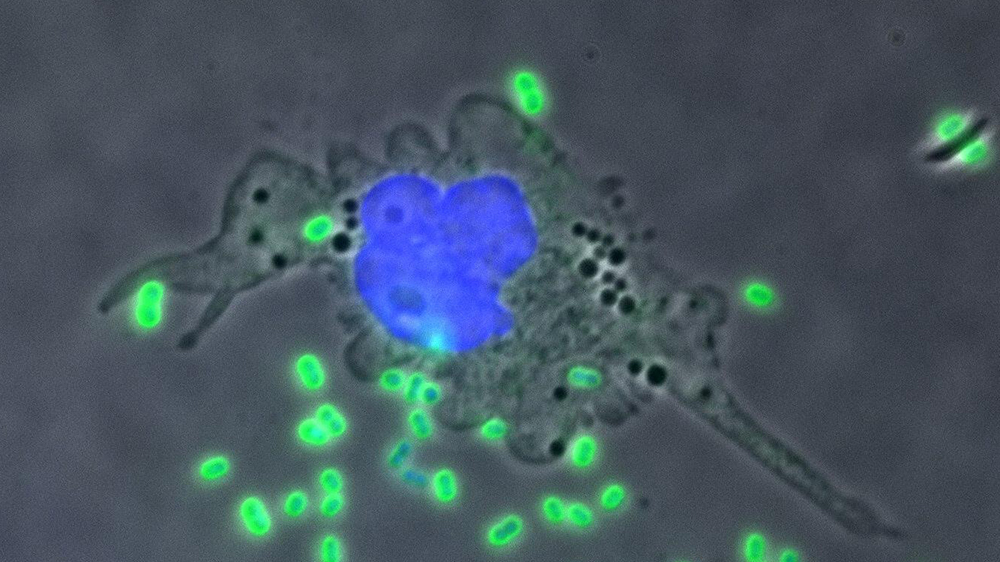
Photographer – Daniel Czyz, University of Florida
A THP-1 cell (ATCC TIB-202) attacking antibiotic-resistant Acinetobacter baumannii.
Photographer – Omer Aras, Memorial Sloan Kettering Cancer Center
Curcumin BF2 accumulation in SK-OV-3 cells (ATCC HTB-77)
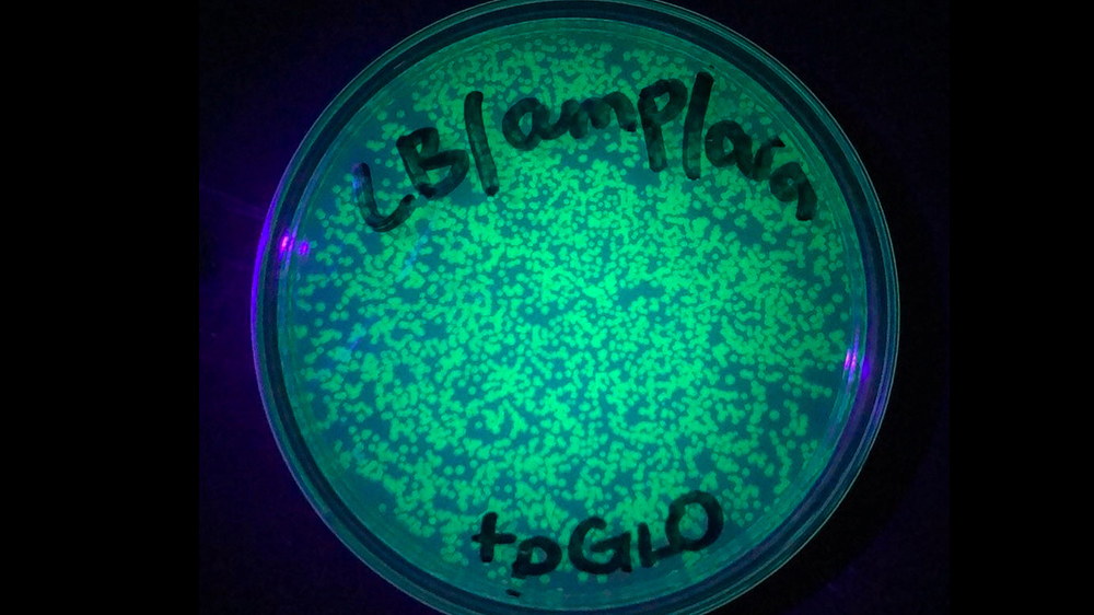
Photographer – Jacob Roy, Plano East Senior High
Transformation of the pGLO plasmid (which includes Green Fluorescent Protein, or GFP).
Photographer – Goutham Kodakandla, Cooper Medical School of Rowan University
S-Acylation of Orai1 promotes CRAC channel formation and its targeting to lipid rafts. HEK-293 (ATCC CRL-1573) Orai TKO cells were transfected with Orai1-GFP and STIM1-RFP and were imaged using TIRF microscopy. Time-lapse TIRF images were obtained after inducing ER store depletion using Thapsigargin. The single images are stacked to create a single image showing the movement of Orai1 and STIM1 to form puncta to promote store-operated calcium entry.
Photographer - Hee-Sung Chae, The University of Mississippi
Differentiated mouse myoblast (C2C12) cells (ATCC CRL-1772) co-stained with a nucleus tracker Hoechst 33342 (blue) and a fluorescent glucose analog 2-NBDG (green).
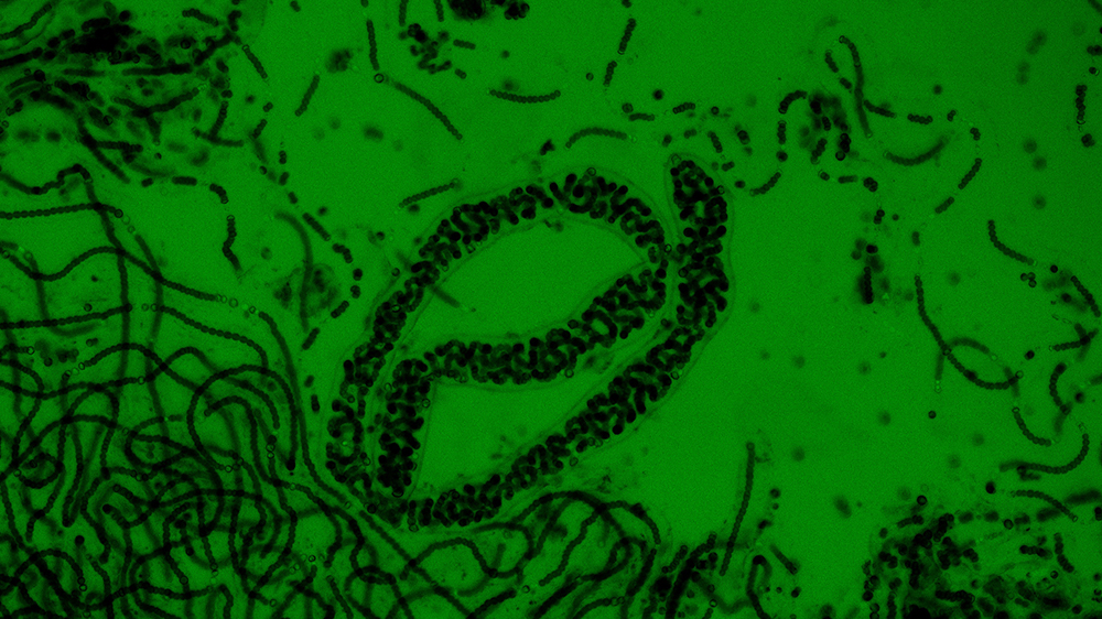
Photographer – Quinne Murphy, University of North Carolina
Phase contrast image of Nostoc sp. (ATCC 27893) at an excitation of 495 nm. Careful inspection shows the presence of heterocysts which allow for the fixation of nitrogen.
Photographer – Jeanette Black, University of North Carolina at Wilmington
Plaque assay showing EHV-1 infecting rabbit kidney epithelial cells (RK-13)
Photographer – Cheyenne Ernst, St. John Fisher College
This photo visualizes leukemia cells (ATCC CRL-9589) in suspension, but uniquely all on the same plane. It is generally difficult to see the cells this was as they are suspended, and some appear closer/farther away, making magnification choice difficult.
Photographer – Adriana LaGier, Grand View University
Cultured HeLa cells (ATCC CCL-2) straining for 5 minutes against vibration produced by an indoor air handler (8.3 ± 0.1 mm/s). Cell stained for actin with phalloidin-AlexaFluor488.
Photograph – Stana Wres, Harvard University
This MEF cell (ATCC SCRC-1040) was fixed with 4% formaldehyde in PBS. Actin (red) is stained with phalloidin-TRITC. Microtubule (blue) and Vimentin (green) are stained with antibodies. Cells were imaged by structure illumination microscopy.
Photographer – Fiona Freeman, Bringham and Women's Hospital
Human MSCs (ATCC PCS-500-012) Loaded with PBAE nanoparticles.
Photographer – Juhyeon Lim< University of Southern California
Mycobacterium tuberculosis H37Rv (ATCC 25618) infects mouse lymphatic endothelial cells (LECs). Using flour microscopy, we observed that GFP-Mtb were internalized.
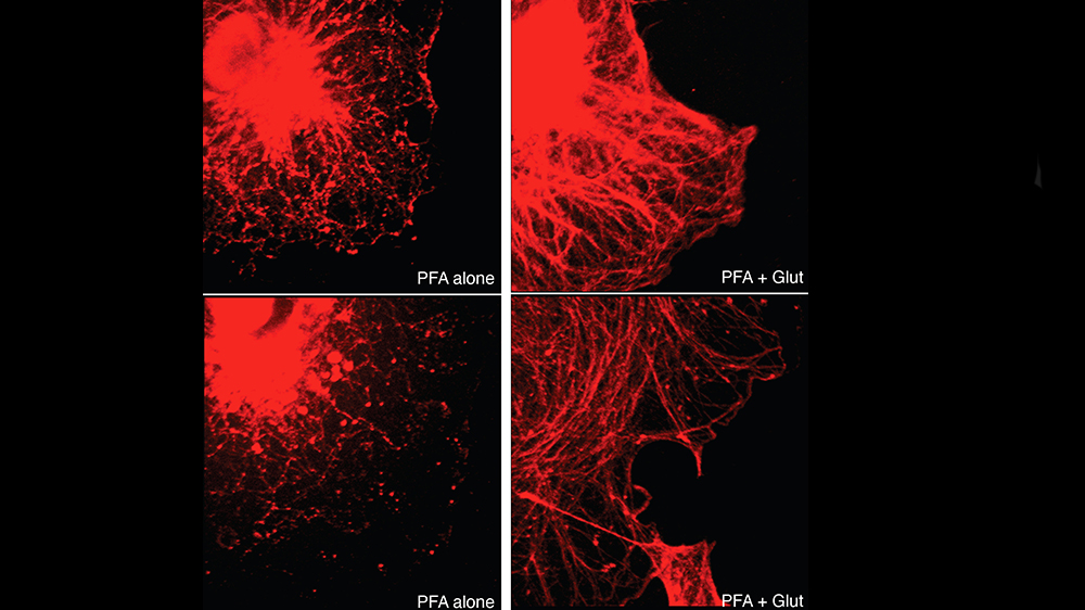
Photographer – Gerado Morfini, University of Illinois
COS-7 (ATCC CRL-1651) were fixed with paraformaldehyde (PFA) in PBS (left panels) or with a mixture of PFA plus glutaraldehyde under microtubule-stabilizing conditions (PFA+Glut; right panels). Cells were then processed for immunofluorescence using anti-tubulin antibodies. Note the remarkable preservation of microtubules in cells fixed with PFA+Glut, compared to cells fixed with PFA diluted in PBS.
Photographer – Rahul Nelli, Iowa State University, College of Veterinary Medicine
Cells: Primary Bronchial/Tracheal Epithelial Cells; Normal, Human (ATCC PCS-300-010)
Stain: NucBlue fixed cell ReadyProbes reagent with DAPI
Scale bar: 100uM
Treatment: mock-infected human tracheal epithelial cells showing nuclear staining.
Comment: We ordered normal human tracheal epithelial cells from ATCC, instead they must have sent cells from Moai from Easter Island.
Photographer – Maodie Wang, Louisiana State University
Human HEp2 cells (ATCC CCL-23) co-stained with a nucleus tracker Hoechst 33342 (blue) and a BODIPY dye (green).
Photographer – Guanyu Zhang, Louisiana State University
Human HEp2 cells (ATCC CCL-23) were co-stained with Golgi tracker (green) and a novel chlorin conjugate (red). The overlay of both shows an orange/yellow color.
Photographer – Taylor Ticer, Medical University of South Carolina
RAW264.7 cells (ATCC TIB-71) stained for nuclei, actin, and a-tubulin.
Photographer – Ritesh Srivastava, University of Alabama at Birmingham
Phase-contrast image of Human Rhabdomyosarcoma (RD) cells (ATCC CCL-136) treated with PP242 (10 µM) for 12 hours.
Photographer – Charles Thirkill, UC Davis
Example of with the immunologic The abnormality of RPE (ATCC CRL-2302) hypersensitivity revealing patient's serum antibody activity on cultured human RPE cells, visualized by indirect immunofluorescence. Antibody activity is confined primarily to the cytoplasm, to the exclusion of the nuclei.

Photographer – Chris Tison, Luna Innovations
Fluorescent micrograph of liposome uptake by S42 cells (ATCC CRL-2942). This material is based upon work supported by the Army SBIR Program/US Army Medical Research and Development Command/U.S. Army Medical Research Acquisition Activity under Contract W81XWH-18-C-0004.
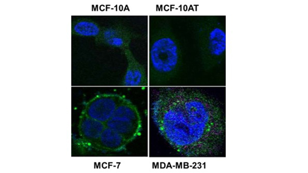
Photographer – Feng Wang-Johanning, SUnnyBay Biotech Inc
The expression of HERV-K was detected in both of breast cancer cell lines (ATCC HTB-22, ATCC HTB-26), but not in non-tumorigenic breast cells (MCF-10A or MCF-10AT) (ATCC CRL-10317) using anti-HERV-K antibody.
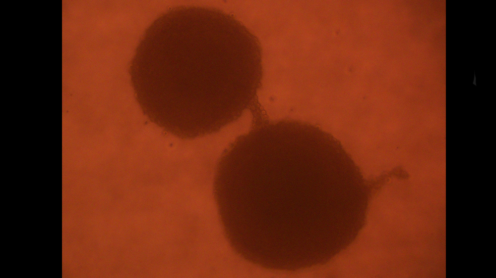
Photographer – Janine Wereley, Medical College of Wisconsin
The human glioblastoma U87 MG cell line (ATCC HTB-14) was grown as a single cell culture in a hanging drop to allow 3-D growth. The resultant neurospheres appear to be communicating with each other via a tubular-like structure composed of cells.
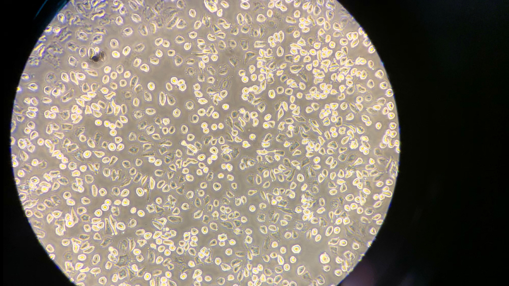
Photographer – Cheyenne Ernst, St. John Fisher College
A549 cells (ATCC CCL-185) in culture following the trypsinization process, which explains why no adherence is seen.
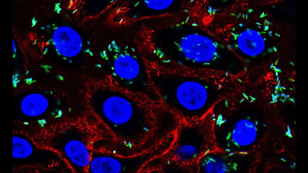
Photographer – Kavita Agarwal, University of California San Diego
Sialidase treated VK2 cells (ATCC CRL-2616), stained with PNA lectin and infected with Fusobacterium nucleatum subsp. nucleatum (ATCC 23726). Blue = DAPI, Red=PNA lectin, Green = F. nucleatum.
Photographer – Mark Parcells, University of Delaware
Zika virus (ATCC VR-1843) infection of Mocha cell (rat cochlear microglial cells line). Fixed and stained with human anti-Zika and goat anti-Human Ig Alexa 488. Nuclei are stained with DAPI.
Past winners
2020 Cell biology winner
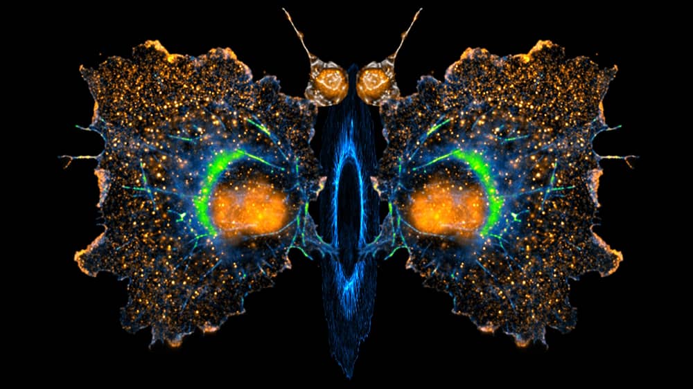
Photographer - Tejeshwar Rao, University of Alabama at Birmingham
Bioart made from three different Cos-7 cells (ATCC CRL-1651) (body, wings and antennas) plated on an RGD coated surface. The organized actin cytoskeleton is displayed with the blue-green or blue LUTs and the paxillin distribution with the orange hot LUT. The cells show differential actin organization and morphologies based on the stiffness of the underlying substrate.
2020 Microbiology winner
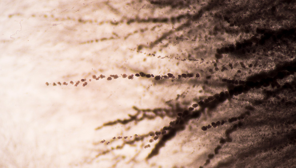
Photographer - Nicanor Austriaco, Providence College
A Yeast Forest" Microscopic image of the edge of a Candida albicans colony (ATCC MYA-2876), a dimorphic yeast that can live either as blastospores or as hyphae. We have shown that the hyphae are more resistant to programmed cell death than their blastophore counterparts.
2020 Cell biology popular winner
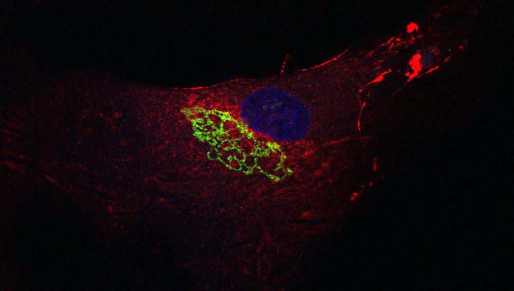
Photographer - Stylianos Z. Karoulias, Icahn School of Medicine at Mount Sinai
The beauty of Golgi Apparatus of Human Dermal Fibroblasts (ATCC PCS-201-012) as it is packaging and preparing Fibronectin for its secretion to the world.
2020 Microbiology popular winner
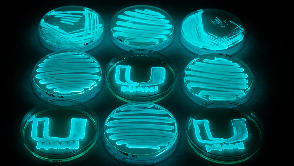
Photographer - Sherwin Reyes, University of Miami
"All about the U: A Glowing Future in Infectious Disease Diagnosis" This is a culture of Photobacterium leiognathi (ATCC 33981) on Trypticase Soy Yeast Extract Salt Water Agar. This glowing bacteria was utilized to make a biosensor to detect pathogens in the urine.