
Authors: Cara N. Wilder, PhD, and Yvonne Reid, PhD
Mycoplasma contamination constitutes a serious concern for cell culturists as these bacterial strains are a common cause of cell line contamination affecting roughly 15-35% of cell cultures and endangering almost all aspects of cell physiology.1,2 This is of particular concern for research laboratories and commercial facilities that employ cell lines in the development and manufacture of cell-derived biopharmaceutical products for medical use.3-7 Contamination of cell substrates used in the production of biopharmaceuticals poses a potential safety risk for patients and presents a serious economic risk for manufacturers in the event of batch adulteration or a product recall. To minimize these risks, routine testing for mycoplasma is performed throughout the product manufacturing and development process. In this article, we will discuss the effects of mycoplasma contamination, how this form of adulteration can affect cell-based drug development, and several quality control techniques and related products that can be used in the detection of mycoplasma contamination.
Mycoplasma are a distinct group of bacterial strains taxonomically ordered under the class Mollicutes. Presently, over 190 species of mycoplasma distributed among humans, animals, insects, and plants are known; of these, only 8 are responsible for approximately 95% of cell culture contamination events.8-10 These microorganisms are distinguishable from other bacterial species by their complete lack of a cell wall, which contributes to the accession of nutrients from a host cell via cytoplasmic exchange. Mycoplasma are also known to be one of the smallest free-living forms of bacteria, ranging in size from 0.15 to 0.3 micrometers.11 This small size enables mycoplasma strains to escape a number of filtration systems as well as grow to a high concentration in cell culture without resulting in media turbidity or other obvious symptoms. In addition to their small size and lack of a cell wall, mycoplasma strains are characterized by a small genome, which drastically reduces their biosynthetic capabilities, causing them to heavily rely on an exogenous source of cholesterol, amino acids, fatty acids, vitamins, and other catabolites provided by their host or environment. If left undetected, mycoplasma contamination can lead to a number of deleterious effects that affect the physiology of cell lines. These include the induction of chromosomal abnormalities, the disruption of DNA and RNA synthesis, changes in membrane antigenicity, the inhibition of cell proliferation and metabolism due to nutrient withdrawal, decreased transfection rates, changes in gene expression profiles, and cell death.1,8,12
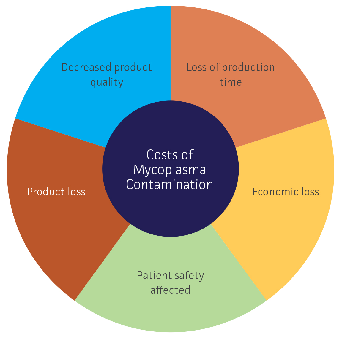
For years, cell lines have been used to produce a number of biological products for therapeutic or medicinal use, including cytokines, viral vaccines, monoclonal antibodies, growth factors, and immunologic modulators. Because of the extensive effects mycoplasma strains have on cell line physiology, metabolism, and gene expression, mycoplasma contamination of cell lines used in the biopharmaceutical industry presents a significant safety risk and economic concern. For the production of biopharmaceuticals, mycoplasma contamination can result in decreased production and may affect the quality of the product. Further, if the mycoplasma contaminant is present in the final biological product, it can directly affect patient safety through the potential to cause disease.11,13 On an economic level, mycoplasma contamination can result in significant costs in the time and expense associated with the loss of impacted batches, investigation into the source of contamination, and decontamination of the facility.13 In turn, this can result in the loss of months of invaluable production time and tens or hundreds of thousands of dollars in associated expenses.14
| Direct agar culture | Indirect Hoechst DNA staining | PCR-based testing | |
|---|---|---|---|
| Advantages | Sensitive Easy to perform Detects viable cells, indicating an active infection |
Rapid Cost-effective Easy to perform Enables the detection of non-culturable mycoplasma strains |
Rapid Reproducible Sensitive Can detect a broad range of mycoplasma species, including non-culturable strains Cost-effective |
| Disadvantages | Laborious Time consuming (28 days) May require expert interpretation Requires selective broth and agar media Not all mycoplasma strains can be easily cultured |
General DNA stain not specific to mycoplasma May require expert interpretation |
Can’t distinguish between viable and non-viable
cells Requires optimization For this method to be implemented, it must show equivalency or superiority to the approved testing procedures |
To mitigate the risk of mycoplasma contamination, microbiological monitoring is required throughout the manufacture of biologicals produced in cell substrates.13,15-18 Some of the most frequently used detection methods include direct agar culture, indirect Hoechst DNA staining, and PCR-based testing. For the production of biopharmaceuticals, the recommended protocols for mycoplasma testing typically rely on the use of culture-based approaches that assess the presence of viable cells in broth, agar, and indicator cell lines.13 However, this form of testing is fairly laborious and requires up to 28 days for completion. This extensive time commitment presents a problem in that some products may have short half-lives whereas other intermediate products may need to be processed quickly. Further, the sensitivity of the culture assay may be affected by the quality of the media components, inconsistencies in media preparation, handling of the mycoplasma culture, interpretation of results, or the lack of mycoplasma reference standards.13
An alternative approach to identifying mycoplasma contamination is through PCR-based testing, which has proven to be a rapid and reliable alternative when validated as a comparable method of detection. This molecular-based method is ideal for research laboratories as it is easy and quick to set-up and analyze. Further, it is highly sensitive, specific, reliable, and fairly cost-effective.13 However, it must be noted that the primary drawbacks to this approach are the inability to distinguish viable from non-viable mycoplasma when targeting genomic DNA and the limited number of primers used. Moreover, without proper assay optimization or the use of appropriate reaction controls, PCR-based methods can be susceptible to false-positive and false-negative results. Currently, many PCR-based methods are designed to amplify the conserved 16S rRNA region of the mycoplasma genome. To ensure the specificity of this method, primers that are broad enough to recognize Mycoplasma, Ureaplasma, Spiroplasma, and Acholeplasma species, as well as specific enough to prevent the amplification of 16S rRNA gene sequences belonging to other non-mycoplasma bacterial contaminants, are essential. For example, the ATCC Universal Mycoplasma Detection Kit (ATCC 30-1012K) enables mycoplasma detection over a wide range of species through the use of universal primers specific to the 16S rRNA gene combined with a touchdown PCR approach. This strategy employs a high annealing temperature in the initial cycle that decreases with subsequent cycles to increase the likelihood of primers binding to the specific targets, reducing the chance that non-specific targets will be amplified. In this case, mycoplasma contamination is easily recognized as a distinct PCR product ranging in size from 434 to 468 base pairs.
Mycoplasma testing resources
Build your assay
Developing your own PCR-based assay? Get started with titered mycoplasma strains and quantitative DNA from ATCC. These mycoplasma positive controls represent species that are most frequently associated with cell culture contamination.
Send us your samples
Don’t have time to test your cell lines? Let the experts in mycoplasma detection handle it. We offer a PCR-based Mycoplasma Testing Service that can quickly and reliably detect over 60 species.
Because the recommended “gold-standard” mycoplasma testing protocol for biopharmaceutical companies requires the use of a culture-based approach to detect viable mycoplasma cells, the implementation of an alternative method, such as the PCR-based approach, must show equivalency or superiority to the approved testing procedures, particularly with regard to the limit of detection.19 However, comparing the limit of detection of a nucleic acid-based testing method to a culture-based approach poses significant challenges with regard to a direct comparison of differing biological measurements. For an impartial assessment of these methods, the use of well-characterized mycoplasma reference materials that represent common cell culture contaminants while demonstrating a high percentage of viable cells and a low degree of aggregation has been recommended.13,19 Because molecular approaches typically rely on the detection of genomic identifiers from mycoplasma strains, regardless of cell viability, the presence of excessive amounts of dead or aggregated material may result in the overestimation of sensitivity.13,19 Thus, the presence of a significant amount of dead or aggregated cellular material can skew the estimated limit of detection of a nucleic acid-based approach. These features can be assessed for reference strains though evaluating the ratio of genomic copies (GC) to colony forming units (CFU). Here, mycoplasma reference strains exhibiting the lowest possible GC/CFU ratio would be indicative of high cell viability and a low degree of aggregation.13
Because developing and implementing a novel PCR-based mycoplasma detection system can be challenging and time consuming with regard to sample preparation and validation of the system to ensure equivalency or superiority to conventional test methods, ATCC has developed the Titered Mycoplasma Reference Strains Panel (ATCC MP-7) for comparing PCR and culture-based detection methods. This panel comprises ten species known to represent common contaminants of cell substrates used in the manufacture of biological products, and was confirmed to have a low GC/CFU ratio and was optimized for high-viability upon thawing. In addition to these products, ATCC also offers quantitative mycoplasma DNA certified reference materials for use as controls in inclusivity/exclusivity testing and establishing limits of detection. These products were derived from the strains comprising the Titered Mycoplasma Strains Panel, and were produced under an ISO 17034 accredited process to confirm identity, well-defined characteristics, and an established chain of custody. Together, these products represent a unique collection of species that are commonly associated with 95% of all mycoplasma contamination in cell culture and are ideal for the validation and comparison of test methods.
Overall, mycoplasma contamination is a major concern for biopharmaceutical producers as it represents a potential safety hazard and economic risk. Routinely testing for contamination throughout the manufacturing process through culture- or molecular-based detection methods can help minimize these risks and may pinpoint any potential sources of contamination. To aid in the development or evaluation of mycoplasma quality control procedures, validated titered mycoplasma reference strains and quantitative mycoplasma DNA reference materials can be used. These products are ideal for comparing test methods and can be used as external controls in evaluating method sensitivity and specificity.
Download a PDF of this white paper
Download NowReferences
- Drexler HG, Uphoff CC. Mycoplasma contamination of cell cultures: Incidence, sources, effects, detection, elimination, prevention. Cytotechnology 39: 75-90, 2002.
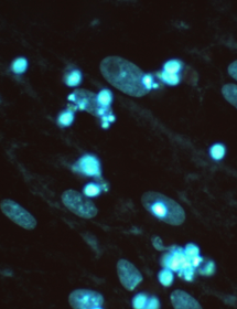
- McGarrity G, Steiner T, Vanaman V. Detection of Mycoplasmal Infection of Cell Cultures by DNA Fluorochrome Staining. Vol. 2, Academic Press, 1983.
- FDA. Innovation or Stagnation: Challenge and Opportunities on the Critical Path to New Medical Products.
- Costa A, Sarmento B, Seabra V. An evaluation of the latest in vitro tools for drug metabolism studies. Expert opinion on drug metabolism & toxicology 10: 103-119, 2014.
- Nayoung K, Ningning H, Sukjoon Y. Cell line modeling for systems medicine in cancers. Int J Oncol 44: 371-376, 2014.
- Zhang D, Luo G, Ding X, Lu C. Preclinical experimental models of drug metabolism and disposition in drug discovery and development. Acta Pharmaceutica Sinica B 2: 549-561, 2012.
- Ozturk S, Hu W. Cell Culture Technology for Pharmaceutical and Cell-Based Therapies. Taylor & Francis Group, LLC, 2005.
- Rottern S. Interaction of Mycoplasma with Host Cells. Physiol Rev 83: 15, 2003.
- Barile MF. Mycoplasmal flora of simians. J Infect Dis 127:S17-20, 1973.
- Bolske G. Survey of Mycoplasma infections in cell cultures and a comparison of detection methods. Zentralbl Bakteriol Mikrobiol Hyg A 269: 331-340, 1988.
- Razin S, Yogev D, Naot Y. Molecular biology and pathogenicity of mycoplasmas. Microbiol Mol Biol Rev 62: 1094-1156, 1998.
- McGarrity GJ, Constantopoulos G, Barranger JA. Effect of mycoplasma infection on pyruvate dehydrogenase complex activity of normal and pyruvate dehydrogenase complex-deficient fibroblasts. Exp Cell Res 151: 557-562, 1984.
- Volokhov DV, Graham LJ, Brorson KA, Chizhikov VE. Mycoplasma testing of cell substrates and biologics: Review of alternative non-microbiological techniques. Molecular and cellular probes 25: 69-77, 2011.
- Champagne D. Advanced Detection of Mycoplasmas - How real-time PCR analysis can save time and money [White paper]. Contract Pharma, 2012. 15.
- EP.6.1 European Pharmacopoeia 6.1., 2008.
- FDA. Center for Biologics Evaluation and Research. Points to consider in “Characterization of cell lines used to produce biologicals. Rockville, MD: US Department of Health and Human Services, 1993.
- FDA. Center for Biologics Evaluation and Research. Guidance for Industry: “Characterization and Qualitification of Cell Substrates and Other Biological Materials Used in the Production of Viral Vaccines for Infectious Disease Indications.” Rockville, MD: US Department of Health and Human Services, 2010.
- ICH. Q5D: Derivation and characterization of cell substrates used for production of biotechnological/biological products. Geneva, Switzerland: International Committee on Harmonization, 1997.
- Dabrazhynetskaya A. et al. Collaborative study report: evaluation of the ATCC experimental mycoplasma reference strains panel prepared for comparison of NAT-based and conventional mycoplasma detection methods. Biologicals : journal of the International Association of Biological Standardization 41: 377-383, 2013.