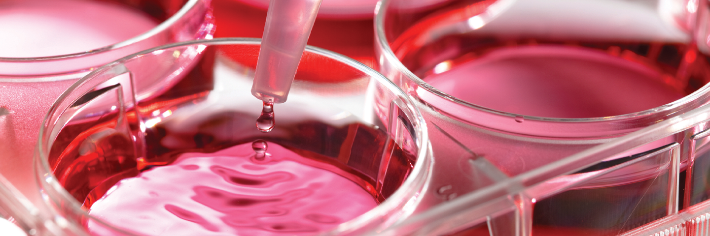
Subculturing, or passaging, is the process by which a population of cells are divided and then transferred into new growth vessels. This process is a critical part of banking and ensures the propagation of the cell line.
Best Practices for Cell Culture
In this webinar, we tap into that vast experience and share the best practices for culturing cells that ensure optimal results and performance. The information delivered in this webinar covers all aspects of successful cell culture, including culture initiation, expansion, authentication, and cryopreservation.
Watch the videoSubculturing Monolayer Cells
Anchorage-dependent cell lines growing in monolayers need to be subcultured at regular intervals to maintain them in exponential growth. When the cells are near the end of exponential growth (roughly 70% to 90% confluent), they are ready to be subcultured. The subculturing procedure, including recommended split-ratios and medium replenishment (feeding) schedules, is specific for each individual cell line.
Subcultivation of monolayers involves the breakage of both intercellular and intracellular cell-to-surface bonds. For some cells that are loosely attached, a sharp blow with the palm of your hand against the side of the flask can dislodge them. Many require the digestion of their protein attachment bonds with proteolytic enzymes such as trypsin/EDTA. For some cell lines mechanical forces such as scraping to dislodge the cells is preferred. After the cells have been dissociated and dispersed into a single-cell suspension, they are diluted to the appropriate concentration and transferred into fresh culture vessels with the appropriate growth medium where they will reattach, grow and divide.
STR Profiling for Mouse Cell Lines
While the authentication of human cell lines has been addressed with STR profiling, up until now the validation of mouse cell lines has been limited at the species level. Watch our on-demand webinar to explore the development of a new STR profiling method for the authentication of mouse cell lines.
Watch the WebinarDon't Risk Your Data
Misidentified and contaminated cell lines undermine your experimental results and discredit preclinical studies. Your research is too important to risk. Be a part of the movement to raise credibility in science and order your cell line authentication and mycoplasma testing services today.
Check your cellsSubculturing Suspension Cells
Most primary cultures, finite cell lines, and continuous cell lines are anchorage dependent and thus grow in monolayers attached to a surface. Other cells, particularly those derived from hematopoietic or certain tumor tissues, are anchorage independent and grow in suspension.
Cell propagation in suspension has several advantages over propagation in monolayer. Subculturing is a simple matter of dilution. There is little or no growth lag after splitting a suspension culture as there is with a monolayer culture, because there is none of the trauma associated with proteolytic enzyme dispersal. Suspension cultures require less lab space per cell yield, and scale-up is straightforward. Cells can be propagated in bioreactors similar to the fermentors used for yeast or bacteria cultures.
Depending upon the cell type, suspension cultures are seeded at densities from 2 × 10⁴ to 5 × 10⁵ viable cells/mL and can attain densities of 2 × 10⁶ cell/mL. If cells are seeded at too low a density they will go through a lag phase of growth, grow very slowly, or die out completely. If cell densities are allowed to become too high, the cells may exhaust the nutrients in the medium and die abruptly.
Recommended seeding and subculturing densities, media replenishment (feeding) schedules, and medium formulations are unique to most cell types and may need to be established empirically. For more information on subculturing animal cells, and depending on the cell type, consult the Animal Cell Culture Guide, the hTERT-immortalized Primary Cell Culture Guide, the Primary Cell Culture Guide, or the Organoid Culture Guide.
How to Initiate, Expand, and Cryopreserve Organoids
Culturing organoids can be intimidating, as these 3D organotypic models require many additional considerations compared to traditional cell growth techniques. Discover how ATCC scientists have used their expertise to create a video tutorial that contains everything you need to know about the initiation, expansion, and cryopreservation of organoids in embedded 3D culture.
Learn more