Keratinocytes are the most common type of skin cell, as they make up the structural component of the epidermis. Keratinocytes divide in the basal to spinous layer (stratum basal and stratum spinosum, respectively). Throughout their program of differentiation, keratinocytes produce a variety of keratin isoforms (a protein that provides strength to skin, hair, and nails). Various crosslinking proteins such as transglutaminase are also produced in the stratum spinosum and activated on the stratum granulosum during the differentiation process. Mature, differentiated keratinocytes are pushed towards the surface of the skin through these layers by the proliferative action of immature keratinocytes in the underlying layers. While being moved through the stratum granulosum and into the stratum corneum, keratinocyte nuclei dissolve and bioactive lipids are squeezed into the extracellular space. The cells are fully differentiated at the stratum corneum, where they are eventually shed into the environment.
Keratinocytes can be isolated from different locations of the body, such as the lining of the oral cavity (human oral keratinocytes; HOK cells). However, the most utilized keratinocytes in primary cell culture are normal human epidermal keratinocytes (NHEK cells) isolated from neonatal or adult skin, the latter of which is significant for the study of adult diseases such as psoriasis and skin cancer. Keratinocytes cultured in serum-free, low calcium growth medium (keratinocyte SFM) will maintain the cells in an un-differentiated, proliferative state while inhibiting fibroblast outgrowth.
Keratinocytes are useful for scientific applications including the study of growth factor behavior and wound healing. Non-melanoma skin cancers such as basal cell carcinoma and squamous cell carcinoma arise from keratinocytes, which provide the functional epidermal barrier.
ATCC primary keratinocytes have been extensively tested by our research and development laboratory and confirmed to form skin equivalents that mimic the architectural features and behavior of normal skin cells.
ATCC offers a variety of dermal cell types applicable to your toxicity and cancer research applications.
Keratinocytes resources
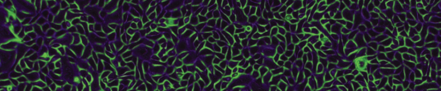
hTERT-immortalized and primary keratinocytes differentiate into epidermal structures in 3-D organotypic culture
Primary and immortalized keratinocyte cultures are essential models for investigating the normal and pathological biology of the epidermis. In this study, we confirmed that the immortalized keratinocyte cell line Ker-CT can differentiate into organotypic skin equivalents in a 3-D culture model.
Read the application note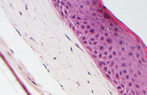
3-D organotypic skin model established using primary keratinocytes and mesenchymal stem cells immortalized by hTERT
In vitro epidermal assay models were established using an ALI Transwell system, primary epidermal keratinocyte cells or Ker-CT, and primary fibroblasts, MSCs, BJ-5ta, or hTERT-MSCs. The study shows that the immortalized keratinocyte co-culture model provides a consistent and robust in vitro system which increases consistency between assays by decreasing the lot to lot variability of primary cells.
See the Research